Medical Tests- FISH
Fluorescent in-situ-hybridization test (FISH)
It is a cytogenetic technique that is used to detect/localize DNA sequences on chromosomes. Fluorescent in-situ Hybridization (FISH) is a big word, to understand it better, let’s break it down.
Fluorescent – fluorescent probes. The fluorescent probes are made from fragments of DNA that were isolated, purified, and amplified for use in the Human Genome Project. These probes bind only to the DNA segment where there is a high degree of sequence similarity. So in the case of CML, they will use probes specifically designed to detect the BCR ABL rearrangement that signals the Philadelphia Chromosome.
in-situ – it is a Latin word that when translated literally means, “in position”. However used in FISH it is referring to the chemical reaction and therefore means: “in the reaction mixture”
Hybridization – as in DNA hybridization, the process of joining two complementary strands of DNA.
So in a sample of your blood, the lab your hospital uses for this test can produce a picture showing exactly how many of your chromosomes are rearranged. At diagnosis it is usually 100%, as you continue to follow your treatments, the ratio of Philadelphia Chromosomes drops. Most patients usually achieve FISH “Undetectable” by six months of treatments (with the newer drugs).
It sounds complicated, but for the patient it requires a simple blood draw.
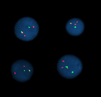
Ever wonder what the FISH picture looks like?

This picture is of inter-phase cells of a CML patient. The secondary colours show the presence of the the translocation which occurs because of BCR ABL, producing the Philadelphia Chromosome.
